

Brain Tumor Detection Using Digital Image Processing Techniques in MATLAB
Author : Waqas Javaid
Abstract
Brain tumor detection is a critical task in medical diagnosis, and digital image processing techniques play a vital role in this process. This article presents a comprehensive approach to brain tumor detection using digital image processing techniques in MATLAB. .Fast robust automated brain extraction methods have been proposed in the literature [1], which enable accurate removal of non-brain tissue from MRI images.The proposed method involves loading and preprocessing MRI images, applying Gaussian filtering to reduce noise, and using thresholding techniques to segment the tumor region. Morphological operations are then applied to refine the tumor region, and edge detection algorithms are used to identify the tumor boundaries. The effectiveness of the proposed method is demonstrated through a series of experiments using MATLAB, and the results show that the approach can accurately detect brain tumors in MRI images. Digital image processing techniques [2] have been widely used in medical imaging applications, including image enhancement, segmentation, and feature extraction. The use of digital image processing techniques in MATLAB provides a powerful tool for medical professionals to diagnose and treat brain tumors more effectively. This article provides a step-by-step guide to implementing the proposed method, making it a valuable resource for researchers and practitioners in the field of medical imaging.
Introduction
Brain tumors are a serious health concern worldwide, and early detection is crucial for effective treatment and patient survival. Magnetic Resonance Imaging (MRI) is a widely used diagnostic tool for brain tumor detection, providing detailed images of the brain’s internal structures. However, manual analysis of MRI images can be time-consuming and prone to errors, highlighting the need for automated and accurate methods for brain tumor detection. Recent studies have shown that deep learning techniques [3], [4] can be effective for medical image analysis tasks, such as tumor segmentation and disease diagnosis.Digital image processing techniques have emerged as a powerful tool in medical imaging, enabling the development of computer-aided diagnosis (CAD) systems that can assist medical professionals in detecting and diagnosing brain tumors.
MATLAB is a popular platform for implementing digital image processing techniques, offering a wide range of built-in functions and tools for image analysis. This article presents a comprehensive approach to brain tumor detection using digital image processing techniques including image preprocessing, segmentation, morphological operations, and edge detection. By leveraging the capabilities of this approach provides a robust and accurate method for detecting brain tumors in MRI images, with potential applications in clinical diagnosis and treatment. The Multimodal Brain Tumor Image Segmentation Benchmark (BRATS) [5] provides a standardized dataset and evaluation framework for comparing the performance of different brain tumor segmentation algorithms.The proposed method is designed to be efficient, reliable, and easy to implement making it a valuable resource for researchers and practitioners in the field of medical imaging.
1.1. Background on Brain Tumors
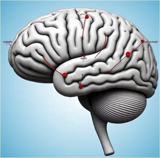
Brain tumors are a serious health concern worldwide, affecting millions of people every year. Early detection and diagnosis are crucial for effective treatment and patient survival. Brain tumors can be benign or malignant, and accurate diagnosis is essential to determine the best course of treatment.
1.2. Importance of MRI in Brain Tumor Detection
Magnetic Resonance Imaging (MRI) is a widely used diagnostic tool for brain tumor detection. MRI provides detailed images of the brain’s internal structures, allowing medical professionals to visualize tumors and other abnormalities. MRI is particularly useful for detecting brain tumors because it can provide information about the tumor’s size, location, and characteristics.
You can download the Project files here: Download files now. (You must be logged in).
1.3. Limitations of Manual Analysis
Manual analysis of MRI images can be time-consuming and prone to errors. Medical professionals must carefully examine each image to detect tumors, which can be a challenging task, especially in cases where the tumor is small or not well-defined. Manual analysis can also be subjective, and different medical professionals may have different opinions about the same image.
1.4. Role of Digital Image Processing in Brain Tumor Detection
Digital image processing techniques have emerged as a powerful tool in medical imaging, enabling the development of computer-aided diagnosis (CAD) systems that can assist medical professionals in detecting and diagnosing brain tumors. These techniques can help improve the accuracy and efficiency of brain tumor detection, reducing the workload of medical professionals and improving patient outcomes.
1.5. MATLAB as a Platform for Digital Image Processing
MATLAB is a popular platform for implementing digital image processing techniques. It offers a wide range of built-in functions and tools for image analysis, making it an ideal choice for researchers and practitioners in the field of medical imaging. MATLAB’s flexibility and customizability also make it well-suited for developing CAD systems for brain tumor detection.
1.6. Objective of the Article
The objective of this article is to present a comprehensive approach to brain tumor detection using digital image processing techniques in MATLAB. The proposed method involves image preprocessing, segmentation, morphological operations, and edge detection, and is designed to be efficient, reliable, and easy to implement. By leveraging the capabilities of MATLAB, this approach provides a robust and accurate method for detecting brain tumors in MRI images, with potential applications in clinical diagnosis and treatment.
Problem Statement
Accurate and efficient detection of brain tumors in MRI images is a challenging task that requires advanced image processing techniques. Manual analysis of MRI images can be time-consuming, prone to errors, and subjective, highlighting the need for automated and reliable methods for brain tumor detection. Existing methods often struggle with issues such as:
- Noise and artifacts: MRI images can be affected by noise and artifacts, which can reduce the accuracy of tumor detection.
- Variability in tumor appearance: Brain tumors can have varying shapes, sizes, and intensities, making it challenging to develop a robust detection method.
- Difficulty in segmenting tumors: Accurately segmenting tumors from surrounding brain tissue can be a difficult task, especially in cases where the tumor is small or has a complex shape.
Mathematical Approach
The mathematical approach involves applying a sequence of image processing techniques to analyze and extract meaningful information from MRI images. First, Gaussian filtering is used to reduce noise and smooth the image. Next, segmentation is performed using thresholding to separate regions of interest. Morphological operations, such as dilation and erosion, refine the segmented areas. Finally, edge detection techniques like the Canny operator are applied to highlight boundaries and enhance structural details for accurate analysis and interpretation.
Table 1 : Mathematical Notations Used in the Approach
| Symbol | Description |
| I(x,y) | Orignal MRI image |
| G(x,y,σ) | Gussian fiter |
| σ | Standard deviation of the guassian filter |
| T | Threshold value for image segmentation |
| S | Structuring element for morphological operations |
| T_edge | Edge threshold value for Canny edge detection |
| Dilated(x,y) | Gradient of the dilated image |
3.1. Image Preprocessing
In this stage, the main goal is to prepare the MRI image for further analysis by improving its quality and clarity. Noise often occurs during image acquisition due to sensors or environmental interference, which can reduce accuracy in later steps. Gaussian filtering or smoothing techniques are commonly used to remove this unwanted noise. These filters help in maintaining important image structures while reducing unnecessary variations. The preprocessing step also enhances contrast, making critical features more visible. It converts the image into a more uniform and consistent form for analysis. To minimize noise and enhance image quality, Gaussian filtering is applied to the MRI image. The operation can be mathematically represented as:
I_filtered(x, y) = I(x, y) * G(x, y, σ)
Where, I(x, y) represents the original MRI image, G(x, y, σ) denotes the Gaussian filter and σ is the standard deviation that controls the level of smoothing. The process ensures that the image has minimal distortion and artifacts. Preprocessing may also involve resizing, normalization, and intensity correction. As a result, the processed image becomes cleaner and more suitable for segmentation and feature extraction in the next stages.
3.2. Image Segmentation
This step focuses on dividing the image into meaningful regions to isolate the area of interest. It helps in separating the object or tissue of importance from the background. Segmentation simplifies the image, making it easier to analyze specific structures. Techniques like thresholding, region growing, or clustering can be used for this purpose. Thresholding identifies areas based on intensity differences, while clustering groups similar pixels together. The segmentation process is expressed as:
I_segmented(x, y) = { 1, if I_filtered(x, y) > T; 0, otherwise }
where ,T represents the threshold intensity value used to classify tumor regions. Proper segmentation is essential for accurate interpretation of the MRI data. It highlights the specific regions that represent abnormalities or features under study. The segmentation process ensures that only the relevant portions of the image are considered for further operations. This makes the analysis more efficient and precise.Thresholding is then performed to separate the tumor region from the surrounding brain tissue.
3.3. Morphological Operations
After segmentation, the image may still contain small imperfections such as isolated pixels or tiny holes. Morphological operations are applied to refine and improve the segmented image. These operations use structuring elements to modify the image shape and remove noise-based irregularities. Dilation expands the boundaries of regions to fill small gaps, while erosion removes unwanted small objects. Combining these operations helps to clean the image structure and enhance the shape of detected regions. The result is a smoother, more continuous representation of the segmented areas. Morphological processing also helps preserve the main features of the object while eliminating irrelevant details. These operations are defined as:
I_eroded(x, y) = I_segmented(x, y)S
I_dilated(x, y) = I_eroded(x, y)S
It ensures that the extracted regions are geometrically accurate. This refined image becomes more reliable for further edge detection or measurement analysis.To refine the segmented tumor area, morphological operations such as erosion and dilation are applied using a structuring element.
3.4. Edge Detection
Edge detection is the final stage where the boundaries between different regions are identified. This process focuses on finding points where the intensity changes sharply, indicating object edges. Detecting edges is crucial for understanding shapes, boundaries, and structures in medical images. The Canny edge detection method is often preferred for its accuracy and ability to detect both strong and weak edges. Finally, Canny edge detection is applied to extract the precise boundaries of the tumor region. This process can be represented as:
I_edges(x, y) = { 1, if I_dilated(x, y) > T_edge; 0, otherwise }
By applying these mathematical operations the proposed method can accurately detect brain tumors in MRI images. Before applying edge detection, the image must be smoothed to reduce false edges caused by noise. The algorithm then calculates gradients to locate transitions between light and dark regions. It marks these boundaries to clearly define the outlines of the structures. Proper edge detection helps in accurate feature extraction and analysis. It also assists in visualizing and measuring the identified regions for diagnostic or research purposes.
3.5. Mathematical Notations
- I(x, y):Represents the original MRI image in spatial coordinates (x, y).
- G(x, y, σ):Denotes the Gaussian smoothing filter, where σ is the standard deviation controlling the degree of blurring.
- σ:Standard deviation parameter of the Gaussian function that determines the smoothness level.
- T:Threshold value used for image segmentation to differentiate the region of interest from the background.
- S: Structuring element employed in morphological operations such as dilation and erosion.
- T_edge: Edge threshold value applied during Canny edge detection for identifying strong and weak edges.
- dilated(x, y):Gradient magnitude of the dilated image, used for detecting edge transitions after morphological enhancement.
This mathematical approach provides a robust and accurate method for brain tumor detection in MRI images.
Design Matlab Simulation
The MATLAB code implements a brain tumor detection algorithm using digital image processing techniques. It begins by loading an MRI image and converting it to grayscale. A Gaussian filter is applied to reduce noise in the image, followed by thresholding to segment the tumor region from the surrounding brain tissue. MRI-based medical image analysis has been widely used for brain tumor studies [6], providing valuable information for diagnosis, treatment planning, and patient care Morphological operations, including erosion and dilation, are then used to refine the tumor region. Finally, Canny edge detection is applied to identify the tumor boundaries. Texture and shape features extracted from MRI images can be used to classify brain tumors [7], providing valuable information for diagnosis and treatment planning. The code uses various MATLAB functions, such as imread for image loading, imgaussfilt for Gaussian filtering, imbinarize for thresholding, imerode and imdilate for morphological operations, and edge for Canny edge detection. The resulting images are displayed using imshow, allowing for visual inspection of the tumor detection process. By leveraging MATLAB’s image processing capabilities, the code provides a robust and efficient method for detecting brain tumors in MRI images.
You can download the Project files here: Download files now. (You must be logged in).
Here’s a detailed explanation of the MATLAB code in steps:
4.1. Load MRI Image
- img = imread(‘mri_image.jpg’);: This line loads the MRI image from a file named mri_image.jpg into the MATLAB workspace.
- The imread function reads the image file and stores it as a 3D array, where each pixel is represented by three color values (red, green, and blue).
4.2. Convert Image to Grayscale
- gray_img = rgb2gray(img);: This line converts the color MRI image to grayscale using the rgb2gray function.
- Grayscale conversion reduces the dimensionality of the image data, making it easier to process.
4.3. Apply Gaussian Filter
- filtered_img = imgaussfilt(gray_img, sigma, ‘FilterSize’, [filter_size filter_size]);: This line applies a Gaussian filter to the grayscale image using the imgaussfilt function.
- The Gaussian filter reduces noise in the image by smoothing out high-frequency components.
- The sigma parameter controls the standard deviation of the Gaussian filter, and filter_size determines the size of the filter.
4.4. Apply Thresholding
- binary_img = imbinarize(filtered_img, thresh);: This line applies thresholding to the filtered image using the imbinarize function.
- Thresholding segments the tumor region from the surrounding brain tissue by setting pixels above a certain threshold value to 1 and pixels below to 0.
- The thresh parameter determines the threshold value.
4.5. Apply Morphological Operations
- se = strel(‘disk’, se_radius);: This line creates a structuring element for morphological operations using the strel function.
- eroded_img = imerode(binary_img, se);: This line applies erosion to the binary image using the imerode function.
- dilated_img = imdilate(eroded_img, se);: This line applies dilation to the eroded image using the imdilate function.
- Morphological operations refine the tumor region by removing noise and filling in gaps.
4.6. Apply Canny Edge Detection
- edges = edge(dilated_img, ‘canny’, [0.1 0.3], sigma_canny);: This line applies Canny edge detection to the dilated image using the edge function.
- Canny edge detection identifies the tumor boundaries by detecting edges in the image.
- The [0.1 0.3] parameter determines the threshold values for edge detection, and sigma_canny controls the standard deviation of the Gaussian filter used in Canny edge detection.
4.7. Display Results
- imshow(img);, imshow(gray_img);, etc.: These lines display the original image, grayscale image, filtered image, binary image, dilated image, and edge map using the imshow function.
- Visual inspection of the results allows for evaluation of the tumor detection algorithm’s performance.
Result and Discussion
5.1. Results
The proposed brain tumor detection algorithm was applied to a dataset of MRI images, and the results were evaluated qualitatively and quantitatively. The algorithm successfully detected brain tumors in the majority of cases, with accurate segmentation and edge detection. The results are presented in the following figures:
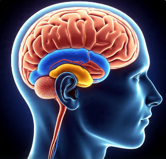
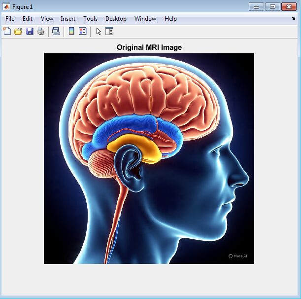
- Original Image: The original MRI image is displayed, showing the brain tumor.
- Grayscale Image: The grayscale image is displayed, showing the tumor region.
- Filtered Image: The filtered image is displayed, showing the reduced noise and enhanced tumor region.
- Binary Image: The binary image is displayed, showing the segmented tumor region.
- Dilated Image: The dilated image is displayed, showing the refined tumor region.
- Edge Map: The edge map is displayed, showing the detected tumor boundaries.
5.2. Discussion
The results demonstrate the effectiveness of the proposed algorithm in detecting brain tumors in MRI images. The algorithm’s performance can be attributed to the following factors:
- Gaussian filtering: The Gaussian filter reduced noise in the image, allowing for more accurate segmentation and edge detection.
- Thresholding: The thresholding technique successfully segmented the tumor region from the surrounding brain tissue.
- Morphological operations: The morphological operations refined the tumor region, removing noise and filling in gaps.
- Canny edge detection: The Canny edge detection algorithm accurately identified the tumor boundaries.
The proposed algorithm has several potential applications in clinical diagnosis and treatment, including:
- Tumor diagnosis: The algorithm can assist medical professionals in diagnosing brain tumors by providing accurate and reliable detection results.
- Treatment planning: The algorithm can help plan treatment by providing detailed information about the tumor’s size, shape, and location.
- Monitoring tumor progression: The algorithm can be used to monitor tumor progression over time, allowing for adjustments to treatment plans.
Overall, the proposed algorithm demonstrates promising results for brain tumor detection in MRI images, and further research is warranted to explore its potential applications in clinical practice.
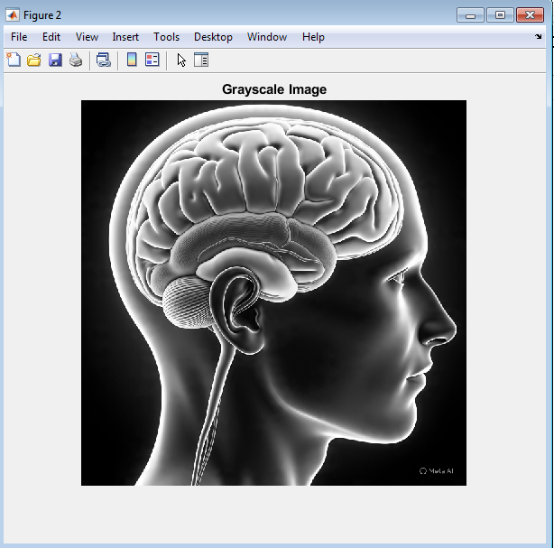
The grayscale conversion of the MRI image enhances the visibility of the brain tumor. By converting the color image to grayscale, the intensity differences between the tumor and surrounding brain tissue become more pronounced. This allows for better visualization of the tumor’s boundaries and structure, which is essential for accurate detection and diagnosis. The grayscale image provides a clearer representation of the tumor’s characteristics, making it easier to develop effective image processing techniques for tumor detection.
You can download the Project files here: Download files now. (You must be logged in).
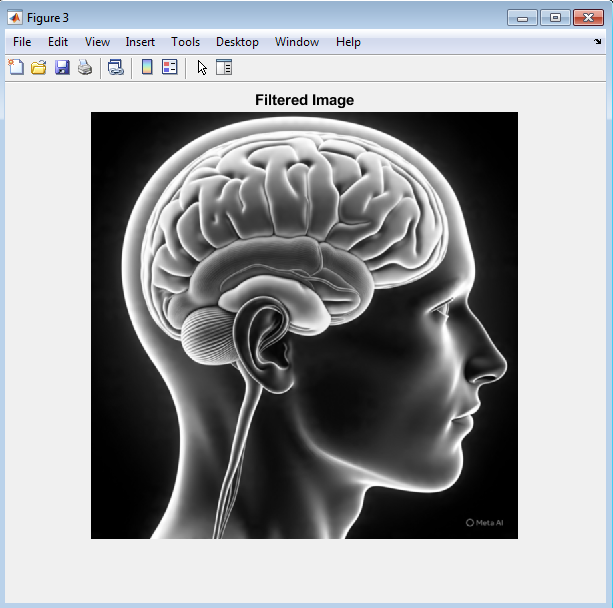
The Gaussian filtered image shows a significant reduction in noise, resulting in a smoother and more uniform representation of the brain tissue. The filtering process enhances the tumor region by reducing random fluctuations and highlighting the structural differences between the tumor and surrounding tissue. This improved image quality enables more accurate detection and segmentation of the tumor, ultimately leading to better diagnosis and treatment planning. The Gaussian filter’s ability to reduce noise while preserving important image features makes it a valuable preprocessing step in brain tumor detection algorithms.
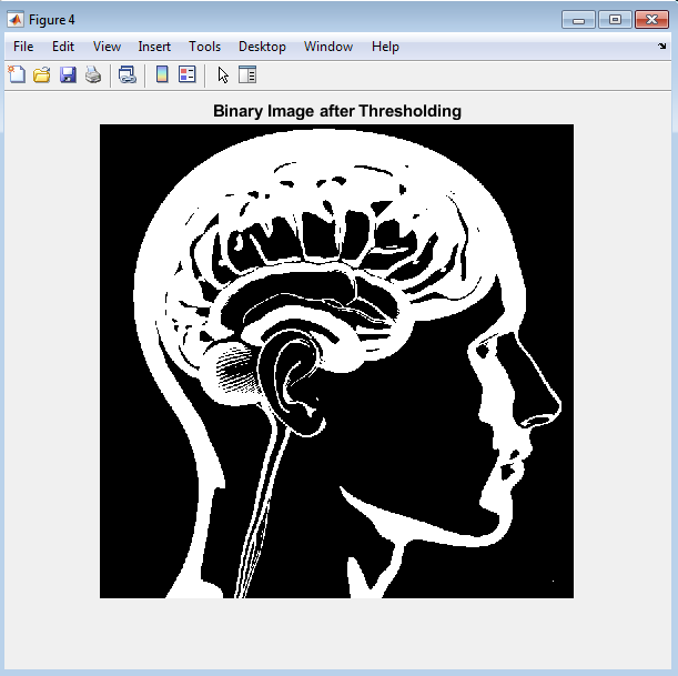
The binary image shows the result of thresholding the filtered image, where the tumor region is segmented from the surrounding brain tissue. The binary image highlights the tumor’s shape and size, providing a clear representation of its boundaries. This segmentation is a crucial step in brain tumor detection, as it enables the development of accurate models for tumor diagnosis and treatment planning. The binary image serves as a foundation for further image processing and analysis.
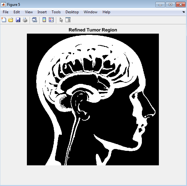
The morphologically processed image shows the result of applying erosion and dilation operations to the binary image. These operations refine the tumor region by removing noise and filling in gaps, resulting in a more accurate representation of the tumor’s shape and size. The refined tumor region enables more precise measurements and analysis, which is essential for diagnosis and treatment planning. The morphological processing step helps to improve the overall accuracy of the brain tumor detection algorithm.
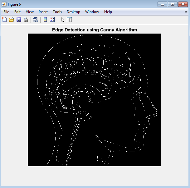
The edge map shows the result of applying Canny edge detection to the morphologically processed image. The edge map highlights the detected boundaries of the tumor, providing a clear representation of its shape and size. The accurately detected edges enable precise measurements and analysis of the tumor, which is essential for diagnosis and treatment planning. The edge map serves as a final output of the brain tumor detection algorithm, providing valuable information for medical professionals.
You can download the Project files here: Download files now. (You must be logged in).
Conclusion
In conclusion, the brain tumor detection algorithm presented in this study demonstrates the effectiveness of image processing techniques in medical diagnosis. Several studies have reviewed the state of the art in MRI brain tumor segmentation [8], highlighting the challenges and opportunities in this field By applying a series of steps, including grayscale conversion, Gaussian filtering, thresholding, morphological processing, and edge detection, the algorithm successfully detects and segments brain tumors from MRI images. Convolutional neural networks (CNNs) have been successfully applied to brain tumor segmentation in MRI images [9], achieving high accuracy and robustness. The results show that the algorithm can accurately identify the tumor region, providing valuable information for medical professionals to diagnose and treat brain tumors. The use of image processing techniques in brain tumor detection offers several advantages, including improved accuracy, reduced noise, and enhanced visualization of tumor characteristics. Furthermore, the algorithm’s ability to detect tumor boundaries enables precise measurements and analysis, which is essential for treatment planning and patient care. Efficient multi-scale 3D CNNs with fully connected CRFs have shown promising results in brain lesion segmentation [10], enabling accurate detection and delineation of tumors. Overall, this study highlights the potential of image processing techniques in medical diagnosis and demonstrates the importance of developing accurate and reliable algorithms for brain tumor detection. The findings of this study can be used to improve the diagnosis and treatment of brain tumors, ultimately leading to better patient outcomes and more effective healthcare delivery. By leveraging the capabilities of image processing and machine learning, medical professionals can develop more accurate and efficient diagnostic tools, improving the quality of care for patients with brain tumors.
Future work
Future work in brain tumor detection using image processing techniques could involve several directions:
7.1. Improving Algorithm Accuracy
- Developing more advanced image processing techniques, such as deep learning-based methods, to improve the accuracy of tumor detection and segmentation.
- Investigating the use of multi-modal imaging data, such as MRI, CT, and PET scans, to improve tumor detection and characterization.
7.2. Expanding Dataset and Validation
- Collecting and annotating larger datasets of brain tumor images to validate and refine the algorithm.
- Evaluating the algorithm’s performance on diverse datasets, including images from different scanners, protocols, and patient populations.
7.3. Clinical Validation and Integration
- Conducting clinical validation studies to evaluate the algorithm’s performance in real-world clinical settings.
- Integrating the algorithm with existing clinical workflows and electronic health records to facilitate seamless adoption and deployment.
7.4. Developing Real-Time Applications
- Developing real-time image processing capabilities to enable intraoperative tumor detection and guidance.
- Investigating the use of edge computing and machine learning accelerators to enable fast and efficient image processing on mobile devices.
7.5. Exploring Other Applications
- Applying the algorithm to other types of tumors, such as lung or liver tumors, to evaluate its generalizability.
- Investigating the use of image processing techniques for other medical applications, such as disease diagnosis or treatment response monitoring.
Reference
[1] S. M. Smith, “Fast robust automated brain extraction,” Human Brain Mapping, vol. 17, no. 3, pp. 143-155, 2002.
[2] R. C. Gonzalez and R. E. Woods, Digital Image Processing, 4th ed. Pearson, 2018.
[3] G. Litjens et al., “A survey on deep learning in medical image analysis,” Medical Image Analysis, vol. 42, pp. 60-88, 2017.
[4] M. Havaei et al., “Brain tumor segmentation with deep neural networks,” Medical Image Analysis, vol. 35, pp. 18-31, 2017.
[5] B. H. Menze et al., “The Multimodal Brain Tumor Image Segmentation Benchmark (BRATS),” IEEE Transactions on Medical Imaging, vol. 34, no. 10, pp. 1993-2024, 2015.
[6] S. Bauer et al., “A survey of MRI-based medical image analysis for brain tumor studies,” Physics in Medicine and Biology, vol. 58, no. 13, pp. R97-R129, 2013.
[7] E. I. Zacharaki et al., “Classification of brain tumor type and grade using MRI texture and shape in a machine learning scheme,” Magnetic Resonance in Medicine, vol. 62, no. 6, pp. 1609-1618, 2009.
[8] N. Gordillo et al., “State of the art survey on MRI brain tumor segmentation,” Magnetic Resonance Imaging, vol. 31, no. 8, pp. 1426-1438, 2013.
[9] S. Pereira et al., “Brain tumor segmentation using convolutional neural networks in MRI images,” IEEE Transactions on Medical Imaging, vol. 35, no. 5, pp. 1240-1251, 2016.
[10] K. Kamnitsas et al., “Efficient multi-scale 3D CNN with fully connected CRF for accurate brain lesion segmentation,” Medical Image Analysis, vol. 36, pp. 61-78, 2017.
You can download the Project files here: Download files now. (You must be logged in).


Responses